Guideme4thesis (GM4T)

Undergraduates

Whipworm – The Hidden Danger in the Gut

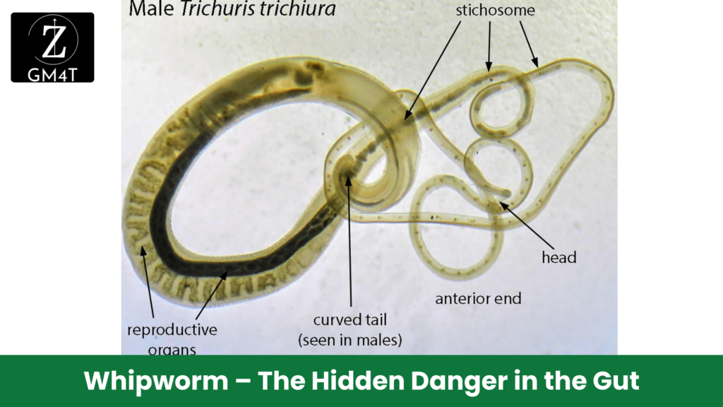
Whipworm infection might sound like something from a horror movie, but it’s a very real and common problem in many parts of the world. Trichuris trichiura, the causative agent, infects the large intestine, often without causing noticeable symptoms in the early stages. But in severe cases, whipworm can lead to chronic diarrhea, anemia, and even rectal prolapse, especially in children. Let’s meet Meera, a 10-year-old girl from a small rural village. Meera had been struggling in school for months, often complaining of a stomachache. Her parents noticed she had lost weight and seemed more tired than usual. When Meera’s condition worsened, they brought her to the local clinic. Stool analysis revealed whipworm eggs, and Meera was diagnosed with trichuriasis. Whipworm infection occurs when individuals ingest whipworm eggs from contaminated soil, usually through unwashed fruits, vegetables, or dirty hands. Once inside the body, the eggs hatch in the small intestine, but the larvae quickly migrate to the large intestine, where they embed themselves in the intestinal lining and mature into adult worms. In mild cases, whipworm infection may go unnoticed, but in severe infections, especially in children like Meera, the impact can be profound. Chronic diarrhea can lead to dehydration and malnutrition, and in some cases, the intestinal lining becomes so damaged that it can lead to rectal prolapse – where part of the rectum protrudes outside the body. Treatment involves antiparasitic drugs like albendazole or mebendazole, which are highly effective. However, prevention is the long-term solution. Improving sanitation, washing hands regularly, and ensuring that fruits and vegetables are thoroughly washed before consumption can significantly reduce the risk of infection.

Whipworm – The Hidden Danger in the Gut Read More »

Hookworm Infection

Hookworm Infection – The Stealthy Blood Thief

Hookworm infection is like a stealthy thief in the night, quietly draining the body of its most vital resource – blood. Caused by parasites such as Necator americanus and Ancylostoma duodenale, this soil-transmitted helminth enters through the skin and eventually settles in the small intestine, where it hooks onto the intestinal wall and begins feeding on blood. Meet Rahul, a 25-year-old farmworker, who had always been energetic and strong. Over the past few months, however, he noticed that he was constantly fatigued, even after a full night’s sleep. His appetite had diminished, and he found it hard to concentrate on his work. When Rahul finally visited the clinic, blood tests revealed severe iron-deficiency anemia. Stool examination confirmed the culprit: hookworm infection. Hookworm larvae live in contaminated soil, waiting for an opportunity to penetrate the skin of an unsuspecting human host. People like Rahul, who walk barefoot in fields or areas where sanitation is poor, are at high risk of infection. Once inside the body, the larvae travel through the bloodstream to the lungs, where they’re coughed up and swallowed, ultimately reaching the small intestine. There, the worms mature and attach themselves to the intestinal wall, feeding on blood. The consequences can be devastating, especially in children, women, and individuals with poor nutrition. Chronic blood loss leads to iron-deficiency anemia, which in turn causes fatigue, weakness, and impaired cognitive development in children. In pregnant women, hookworm infection can result in low birth weight, stillbirth, or complications during childbirth. The good news is that hookworm infection is preventable and treatable. Wearing shoes, improving sanitation, and participating in community deworming programs can significantly reduce infection rates. Early detection and treatment with anti-parasitic drugs like albendazole or mebendazole are key to preventing long-term health complications. Don’t let a hidden blood thief sap your strength. Advocate for better sanitation in your community, participate in deworming programs, and protect yourself and your family from this stealthy parasite.

Hookworm Infection – The Stealthy Blood Thief Read More »

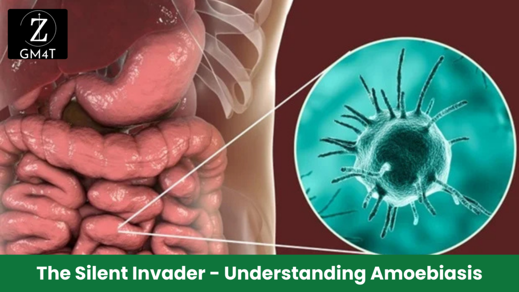
The Silent Invader - Understanding Amoebiasis

The Silent Invader – Understanding Amoebiasis

Unmasking Amoebiasis: The Silent Threat Lurking in Our Water Amoebiasis is like a sneaky intruder – often silent but capable of causing serious damage if left unchecked. It’s caused by a tiny protozoan parasite called Entamoeba histolytica, which primarily invades the human gastrointestinal tract. While the parasite can live peacefully inside many individuals, it has the potential to become aggressive, leading to intestinal or even life-threatening liver infections. Take the case of Raj, a 35-year-old businessman from a small town. Raj had been experiencing mild stomach pain and occasional loose stools for weeks but brushed it off as “just something I ate.” One evening, he developed severe abdominal pain and a high fever. Upon visiting a doctor, imaging revealed a liver abscess – a classic sign of extraintestinal amoebiasis. Raj was shocked when he learned that an infection he had likely contracted from unclean water months ago was now putting his life at risk. In many cases, amoebiasis remains asymptomatic, especially in areas with poor sanitation, where the parasite thrives. Contaminated food, unfiltered water, or contact with dirty surfaces can lead to the ingestion of Entamoeba histolytica cysts. These cysts are resilient, surviving in the harshest conditions outside the human body, only to become active once they reach the intestines. Here, they either remain dormant or invade the intestinal lining, leading to symptoms like diarrhea, stomach cramps, and in severe cases, dysentery with blood and mucus in the stool. But the real danger lies in the potential for the parasite to leave the intestines and travel to other organs, particularly the liver. This is when the disease becomes far more dangerous. If you’re experiencing chronic gastrointestinal discomfort, don’t wait for a crisis like Raj’s. Seek medical advice early, especially if you live in or have traveled to an area where amoebiasis is prevalent. Remember, this sneaky intruder is always looking for the next unsuspecting host. It’s time to take action! Clean water, good sanitation, and early diagnosis are your best defenses against this disease. If you’re in a high-risk area, make sure your food is well-cooked, and avoid untreated water. And if you suspect symptoms of amoebiasis, don’t delay – early treatment can save lives. Read Also :-

The Silent Invader – Understanding Amoebiasis Read More »

"ascariasis symptoms", "ascariasis is caused by", "ascariasis treatment", "ascariasis surgery", "ascariasis pictures", "ascariasis photo"

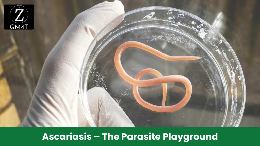
Ascariasis – The Parasite Playground

Imagine your intestines as a bustling playground. In most people, things are in order, but for those affected by ascariasis, the playground becomes overrun with unwelcome visitors. Ascariasis is caused by the roundworm Ascaris lumbricoides, which can grow up to 35 cm. These parasites can cause more than just discomfort; they rob you of essential nutrients and can lead to serious complications if not addressed. Consider Priya, an energetic 7-year-old who suddenly became sluggish and irritable. She had persistent abdominal pain, occasional nausea, and a poor appetite—common ascariasis symptoms. Her parents assumed it was just a passing bug, but when Priya started losing weight and became visibly weak, they rushed her to the doctor. A stool test revealed an infestation of Ascaris lumbricoides, one of the most common soil-transmitted helminths in children, especially in rural areas. Ascariasis is caused by accidentally ingesting the eggs of Ascaris lumbricoides, often through contaminated food or soil. Once inside the body, these eggs hatch into larvae that travel through the bloodstream, making their way to the lungs before being coughed up and swallowed again. Once back in the intestines, they mature into adult worms, where they thrive and lay thousands of eggs daily. While mild infections may go unnoticed, heavy infestations can lead to malnutrition, growth retardation, and intestinal obstruction. Children like Priya, who play in contaminated soil or live in areas with poor sanitation, are particularly vulnerable. The worms not only compete for the food that children eat but may also trigger growth delays and impair cognitive development. If left untreated, these parasites can cause serious complications, and in extreme cases, ascariasis surgery may be required to remove blockages caused by the worms. Fortunately, the ascariasis treatment is simple and effective, typically involving a single dose of albendazole or mebendazole. Visual aids like ascariasis pictures and ascariasis photos can help educate parents about the appearance of these parasites and their potential impact on health. Prevention is the real goal. Good sanitation, regular deworming in endemic areas, and proper food hygiene can protect children from these uninvited visitors in their intestinal playgrounds. Parents and caregivers, it’s time to prioritize deworming, especially in high-risk areas. Protect your child’s health and well-being by ensuring regular health check-ups, good hygiene practices, and participation in deworming programs. Don’t let the playground of life get overrun by parasites!

Ascariasis – The Parasite Playground Read More »

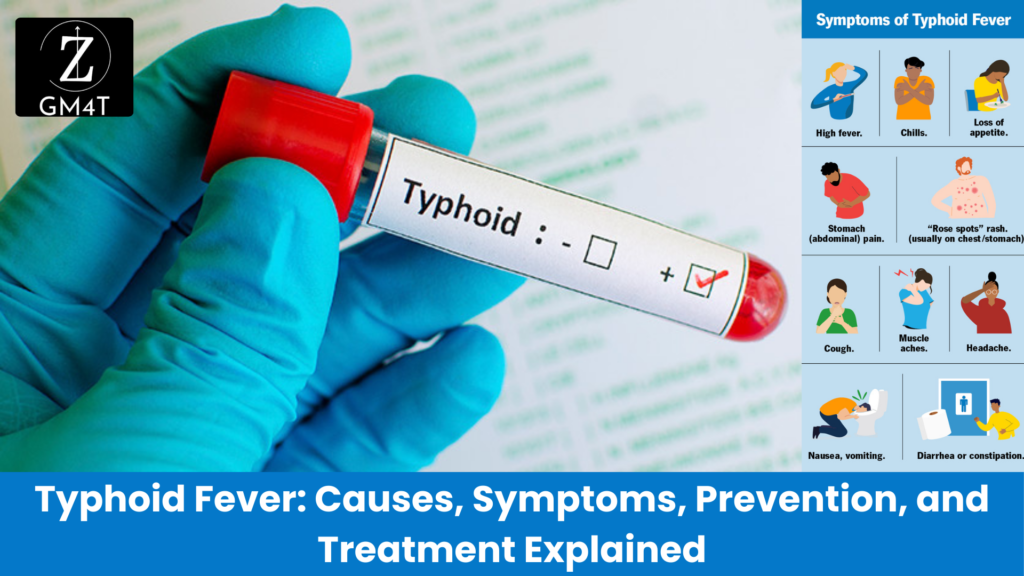
Typhoid Fever: Causes, Symptoms, Prevention, and Treatment Explained

Typhoid Fever: Causes, Symptoms, Prevention, and Treatment Explained

Let me take you back to 2019, to the quiet, semi-urban town of North India. Life was going on as usual, but beneath the surface, a public health disaster was brewing. One morning, a young man visited the local clinic, complaining of high fever, headache, and stomach pain. The doctor suspected it was a viral infection, a routine case. But as the days went by, more and more patients began showing up—each one with the same set of symptoms: fever that just wouldn’t quit, intense stomach pain, and extreme fatigue. Before long, the number of patients swelled to 75, and the town’s health workers realized something much bigger was happening. These weren’t just isolated cases—they were witnessing an outbreak. The Hunt for the Source What could be causing this mysterious illness? The local health department sprang into action, conducting a thorough investigation. A sanitary survey revealed the root of the problem: a leaking sewage pipe had spilled waste into the town’s drinking water supply. The same water people had been drinking, cooking with, and using for daily activities had been tainted with dangerous bacteria. That bacteria was none other than Salmonella typhi, the culprit behind typhoid fever. The town’s water was carrying this tiny organism from person to person, like a stealthy intruder, infecting them with enteric fever. The Symptoms Unfold Typhoid fever isn’t your average illness. It comes in slowly, insidiously— like a thief in the night, slipping through the defenses of the body, unnoticed at first but leaving chaos in its wake. At first, it seems like nothing more than a mild inconvenience—a little fever, a headache, some stomach discomfort. But then it tightens its grip, day by day, rising stealthily like water in a flood. By the time you realize the danger, it’s already overwhelmed you, leaving you feverish, exhausted, and vulnerable to its destructive complications. It’s a silent invader that operates under the radar, but when it strikes, it hits like a hammer, hard and unforgiving. Patients  didn’t know what they were up against. Many suffered from severe abdominal pain, feeling like their insides were being twisted into knots. Some developed coated tongues, a telltale sign of typhoid, as if their mouths were cloaked in sickness. Others had rose spots—pink patches blooming on their skin, like a mark left by the disease itself. For some patients, it wasn’t just a matter of feeling sick. The bacteria had invaded their bloodstream, like an army crossing enemy lines, spreading the infection to the liver, spleen, and gastrointestinal tract, putting them at risk of life-threatening complications like intestinal perforation or hemorrhage. The Turning Point Once the source of infection was identified, the public health officials acted quickly. They repaired the sewage leak and provided clean drinking water to the community. But stopping the outbreak required more than just fixing the pipes. It was a race against time to treat those who were already infected. Doctors administered antibiotics like ceftriaxone and azithromycin to tackle the multidrug-resistant strains of Salmonella typhi, a growing threat in India. The patients received supportive care to manage their fever and rehydration for those suffering from diarrhea or vomiting. It was a fight on two fronts—against the bacteria inside their bodies and the unsanitary conditions outside. Education played a big role too. Health officials went door to door, teaching families about the importance of boiling water, practicing good hygiene, and washing hands. They worked to ensure that even though the outbreak had started with a public health failure, it wouldn’t end with one. The Aftermath and Lessons Learned In the end, no fatalities were reported, thanks to the swift public health response and timely treatment. This outbreak was a wake-up call—not just for the town, but for everyone. It served as a reminder of how typhoid fever is like a hidden predator, waiting for the right conditions—contaminated water, poor sanitation—to pounce. It also showed us that typhoid fever isn’t just a disease of the past. It’s still here, still affecting millions of people, especially in places where water infrastructure is weak, like a crack in a dam, ready to unleash devastation. But this story also gives us hope. It shows that with the right tools—clean water, vaccination, education, and timely medical intervention—typhoid’s grip can be loosened, its spread halted. It’s a story of resilience, like a village rising from the ashes, where public health measures saved the day, proving that even the most insidious diseases can be stopped when communities and healthcare systems work together. Typhoid Fever: Understanding a Silent Predator In 2019, a quiet town faced a health crisis that many thought belonged in the past. Typhoid fever—an illness often overshadowed by other global diseases—reared its head, revealing how vulnerable communities can still be to this ancient foe. But what caused the outbreak, how does typhoid spread, and what can be done to prevent future cases? This is the story of an outbreak, a bacteria, and a battle against time. A Hidden Threat: The Cause of Typhoid Fever Typhoid fever isn’t your average illness. It’s caused by the bacterium Salmonella typhi, a microorganism transmitted through contaminated food and water. Once inside the human body, it invades the gastrointestinal tract, quietly making its way into the bloodstream. Like a stealthy intruder, it spreads unnoticed at first, causing symptoms that seem minor: a headache, slight fever, or stomach discomfort. But this bacteria isn’t here for a short stay. It tightens its grip, day by day, rising like a flood, bringing severe fever, exhaustion, and abdominal pain. When left untreated, it can cause serious complications—like a hammer striking—with the risk of intestinal perforation, hemorrhage, and even death. In developing countries, where access to clean water and sanitation can be limited, typhoid thrives. This is not just a disease of the past. Today, typhoid fever remains a global health concern, As of 2019, an estimated 9 million people get sick from typhoid and 110 000 people die from it every year (World Health Organization). Most cases occur in

Typhoid Fever: Causes, Symptoms, Prevention, and Treatment Explained Read More »

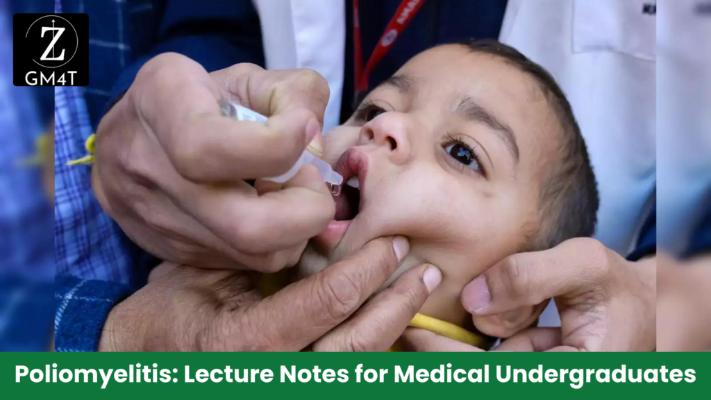
Poliomyelitis: Lecture Notes for Medical Undergraduates

Poliomyelitis Lecture Notes for Medical Undergraduates

About the Disease Discover – Poliomyelitis Lecture Notes for Medical Undergraduates – Poliomyelitis (polio) is a highly infectious viral disease that primarily affects children under 5 years of age. It is transmitted mainly through the faecal-oral route and multiplies in the intestine, from where it can invade the nervous system and cause paralysis. The deficit is purely motor, with sensation intact. Transmission • Person-to-person spread via faecal-oral route. • Contaminated water or food. • Virus multiplies in the intestine and can lead to paralysis. Mnemonic: F-W-C (Faecal-oral, Water, Contaminated food) Epidemiology Source: WHO. Endemic Countries: Pakistan and Afghanistan About the Virus • Three serotypes: Type 1, Type 2, Type 3. • Immunity to one serotype does not confer immunity to the others. • Type 2 eradicated in 2015, last seen in India in 1999. • Type 3 eradicated in 2019, last seen in 2012. • Only Type 1 still circulates today, primarily in two countries. • Global polio incidence has decreased by 99%. Vaccine-Associated Paralytic Poliomyelitis (VAPP) • Caused by loss of viral attenuation in OPV. • Extremely rare • Linked to serotype 3, more common in immunodeficient patients. Mnemonic: R-S-I (Rare, Serotype 3, Immunodeficient) Circulating Vaccine-Derived Poliovirus (cVDPV) • OPV virus can spread in areas with poor sanitation and offer passive immunization. • In under-immunized populations, excreted virus can circulate for an extended time, undergo genetic changes, and mutate into a form that can cause paralysis. • cVDPV takes at least 12 months to emerge in under-immunized populations. • Low vaccination coverage is the main problem, not the vaccine itself. • Solution: 2-3 rounds of high-quality immunization campaigns to stop the virus. Mnemonic: I-P-G-M (Immunodeficient, Prolonged circulation, Genetic mutation, Mutated virus causing paralysis) Diagnosis • Tests include blood, cerebrospinal fluid (CSF), respiratory, and stool viral cultures, as well as PCR to detect poliovirus. Mnemonic: B-C-R-S-P (Blood cultures, CSF, Respiratory cultures, Stool cultures, PCR) Complications • Major complications: Paralysis with bulbar involvement, fatal respiratory and cardiovascular collapse, and postpolio syndrome. • Postpolio syndrome: New-onset or progressive muscle weakness in previously diagnosed polio patients. Mnemonic: P-R-P (Paralysis, Respiratory collapse, Postpolio syndrome) Prevention of Polio AFP Surveillance: 4 steps 1. Finding and Reporting AFP cases: Detect at least 1 case of AFP per 100,000 children under 15 years of age. 2. Transporting Stool Samples: Samples should arrive at the lab within 72 hours, with two specimens collected 24 hours apart and arriving via reverse cold chain. 3. Isolating Poliovirus: Distinguish between wild and vaccine-related poliovirus. 4. Mapping the Virus: Map the genetic makeup and geographical areas of circulation. • Note: 80% of AFP cases should have a follow-up examination at 60 days after onset of paralysis. Mnemonic: F-T-I-M (Finding, Transporting, Isolating, Mapping) Polio Eradication Strategy 2022–2026 strategic framework: Source WHO Tip: Remember the vision and goals. Current Challenges • Afghanistan: Ban on house-to-house immunization has led to over 1 million children being missed, with 90% of WPV1 cases in inaccessible areas. • Poor-quality campaigns due to insufficient planning, staffing issues, and lack of accountability. • Pakistan: Progress stalled due to complacency, leadership transitions, vaccine hesitancy, and misinformation on social media. • Marginalized Pashto-speaking communities disproportionately affected (81% of WPV cases). Mnemonic: M-P-L-V (Missed children, Poor campaigns, Leadership issues, Vulnerable communities) Sources: Practice Questions Multiple Choice Questions (MCQs) Answers to MCQs Subjective Practice Questions Read Also :-

Poliomyelitis Lecture Notes for Medical Undergraduates Read More »

Comprehensive Guide to Measles, Mumps, and Rubella: Symptoms, Diagnosis, and Prevention

Comprehensive Guide to Measles, Mumps, and Rubella: Symptoms, Diagnosis, and Prevention

Discover – Comprehensive Guide to Measles, Mumps, and Rubella: Symptoms, Diagnosis, and Prevention: This Ready Reckoner has been designed to provide medical students with a quick and comprehensive reference for three significant childhood viral infections: Measles, Mumps, and Rubella. As part of their clinical education, students are often tasked with diagnosing and managing these common infections. The reckoner simplifies and condenses the essential clinical features, complications, diagnostic methods, and prevention strategies for each disease, enabling students to quickly review key concepts before exams or clinical rounds. It also serves as an aid in understanding the differential diagnosis of fever with rash in children, a crucial skill in pediatric and infectious disease management. By bridging theory with clinical practice, this resource equips students to recognize these conditions and make informed decisions in real-world scenarios. Measles (Rubeola) Mumps Rubella (German Measles) Key Differences Between Measles, Mumps, and Rubella Feature Measles Mumps Rubella Agent RNA paramyxovirus RNA virus from Rubulavirus genus RNA virus from Togavirus family Transmission Respiratory droplets Respiratory droplets, contact with saliva Respiratory droplets, direct contact Incubation Period 10-14 days 14-18 days 14-21 days Clinical Features High fever, cough, coryza, Koplik’s spots, rash Swelling of parotid glands, fever Mild fever, rash, swollen lymph nodes Complications Pneumonia, encephalitis, SSPE Orchitis, pancreatitis, meningitis CRS in pregnancy, arthralgia Vaccine MMR (Measles, Mumps, Rubella) MMR (Measles, Mumps, Rubella) MMR (Measles, Mumps, Rubella) Prevention 95% vaccination coverage 95% vaccination coverage 95% vaccination coverage Important Points to Remember

Comprehensive Guide to Measles, Mumps, and Rubella: Symptoms, Diagnosis, and Prevention Read More »

Open chat
Hello 👋
Can we help you?